Full text rendered from the published PDF. The PDF is the version of record; if the two differ, the PDF governs.
Cardiovascular disease, and particularly myocardial infarction (MI), is the leading cause of disability and death in women worldwide. Young females with MI have previously been disregarded from inclusion in mass epidemiological research and clinical trials due to the physiologically protective properties of oestrogen. Moreover, young women who present with chest pain are often not considered at a risk for MI, which often delays workup and timely intervention, and they remain an unrecognised and under-treated subgroup. We report a case of a 22-year-old, medically free female with MI, with no traditional risk factors, complicated by a final, unfortunate outcome of death. This case report highlights the need to thoroughly investigate the prevalence, symptoms, and types of acute MI in young female patients, as well to create validated tools to assess the risk factors associated with this subgroup.
Keywords: Acute Myocardial Infarction, Angiography, Female, Young Adult
Myocardial infarction (MI) occurs when blood flow to the coronary artery slows or stops, damaging the cardiac muscle with disastrous health consequences [1]. The leading cause of mortality and morbidity in women worldwide is cardiovascular disease, and MI in particular [2].
While young females with MI have previously been disregarded from inclusion in mass epidemiological research and clinical trials due to the physiologically protective properties of oestrogen, recent data indicates a significant incidence of mortality and morbidity in this group [3]. Additionally, when compared with similarly aged men, young women with heart
A 22-year-old Saudi female, medically and surgically free, presented to our emergency department (ED) with a history of chest pain. The pain, located substernally, started suddenly two hours before arrival, was described as crushing in nature, constant and worsening in severity, radiating to both arms but more to the left, and was
Falwah Alharthi, Abeer Alfadhliah, Rawan Alosaimi are with The Emergency Department, King Abdelaziz Medical City, Riyadh, Saudi Arabia, e-mail: Felwaalharthi@gmail.com, e-mail: Abeerbf5@gmail.com, e-mail: rfalosaimi@KAAUH.edu.sa Ahmed Alharkan is with King Abdullah University Hospital, Riyadh, Saudi Arabia, e-mail: Alharkan@live.com DOI : 10.52609/jmlph.v4i1.107
disease have a higher risk of mortality from acute MI [4]. More specifically, their chance of being readmitted after 30 days following an MI is roughly two-fold higher [5]. Risk factors attributed to MI are not yet well established; however, according to one major case-control study, younger patients were found to have more significant risk factors than older patients with regard to smoking, lipid abnormalities, hypertension, and diabetes [6].
In the clinical presentation of MI, young women are more likely to have atypical symptoms which result in delayed recognition and treatment. In one study, women presented with a greater number of additional non-chest pain symptoms than men, including epigastric abdominal pain, and discomfort in the jaw, neck, and arms [7]. Furthermore, four categories can be used to group the causes of MI in young people: chemical substance abuse, hypercoagulable state, non- atherosclerotic coronary heart disease, and atherosclerotic coronary heart disease [8]. However, coronary artery atherosclerosis remains the leading cause of myocardial infarction in young adults, accounting for approximately 80 percent of cases in this demographic [9].
Given the increased mortality and morbidity associated with premature MI in young women, it is imperative to thoroughly investigate the prevalence, symptoms, and types of acute MI in this patient cohort, and to explore how symptom recognition influences patients’ care-seeking behaviours and early interactions with healthcare providers.
associated with palpitations, nausea, and shortness of breath. The patient reported that the earliest symptom was palpitations that started upon waking the same day with a presyncope attack, after which the chest pain developed. A past history of palpitations and similar milder chest pain was reported, starting six months prior to presentation; the patient had not sought medical advice regarding her symptoms, although she attended family health clinics at our facility for routine laboratory tests and had a normal total cholesterol with normal liver and renal function during her visits. Systemic review was unremarkable. The patient denied smoking and substance or drug abuse. Past family history was remarkable for significant cardiac disease: her father was known for ischaemic heart disease and had undergone seven cardiac catheterisations, while her mother was known for valvular heart disease. There was no history of sudden cardiac death in the family. The patient’s initial measured vital signs were: blood pressure 107/97 mmHg, heart rate 77/min, respiratory rate 15/min, temperature 36.6 ◦ C, and oxygen saturation 98% on room air. Clinically, the patient was conscious, alert, and oriented; appeared in pain and distress with tenderness upon chest wall palpation; her pupils were equally reactive bilaterally; and there was no sign of needle tracks or skin changes. Other physical examinations were unremarkable.
Epinephrine 0.5 mg was administered followed by epinephrine and norepinephrine infusions. After four cycles of CPR, the patient achieved return of spontaneous circulation (ROSC); two minutes later, she arrested again with a pulseless electrical activity (PEA) rhythm, and Advanced Cardiovascular Life Support (ACLS) protocol was re-initiated. The patient achieved ROSC followed by arrest seven times; after the seventh arrest episode she did not achieve ROSC and her subsequent rhythms were persistently ventricular fibrillation. A 200 J to 400 J biphasic shock was delivered in every cycle, and epinephrine 1 mg was given thirty-seven times with multiple doses of epinephrine IV push. She also received amiodarone 150 mg IV, lidocaine 60 mg IV, and magnesium sulphate 2 g IV, while sodium chloride 100 ml was given eight times, calcium chloride 1 g three times, and phenylephrine 400 mcg IV three times. As ROSC had occurred multiple times, the decision was made for extracorporeal membrane oxygenation (ECMO). A local tertiary hospital with ECMO services was contacted and, after approximately four hours of continuous CPR, ECMO was performed and the patient was transferred to the facility for continuity of care. Unfortunately, her clinical status remained critical, and she was pronounced dead the following day. The patient was medically free, neither a smoker nor a substance abuser, had a previously normal lipid profile, and had no traditional risk
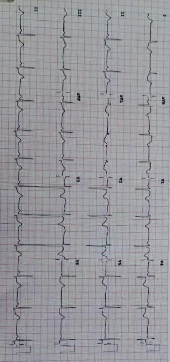
A 12–lead electrocardiogram (ECG) showed an ST-segment elevation of greater than 1 mV in the aVR, with widespread ST depression in the anterior, lateral, and inferior leads (Figure 1). Chest X-ray was unremarkable, with no signs of widened mediastinum, pneumothorax, pneumomediastinum, pneumonia, effusion, or perforation. Laboratory results were within normal limits, except for a mildly elevated troponin level: a value of 21/mL with a normal range of 12.6- 20.7 pg/mL.
The patient received 300 mg aspirin, 300 mg clopidogrel, a 50 mcg fentanyl intravenous push, and a bolus of intravenous fluid in the ED. The cardiology service was consulted early, and the cardiac catheterisation laboratory (cath lab) was activated to manage the case accordingly. Another 300 mg clopidogrel was given with 5000 units of unfractionated heparin, and a nitroglycerin infusion was started at 10 mcg/minute; the patient was then moved immediately to the cath lab. There
, she was initially vitally stable. Findings of cardiac catheterisation showed total occlusion of the left main (LM) coronary short artery, the left anterior descending artery (LAD), and the left circumflex artery (LCx). The right coronary artery (RCA) was normal with collaterals to the left system. Canulation commenced with some difficulty; however, when crossing the LCx the patient went into cardiac arrest. Cardiopulmonary resuscitation (CPR) was started, and a chest compression system machine was attached with continued CPR.
During CPR, the LM and LCx were ballooned and a 2.5x15 drug-eluting stent was inserted into both. A guidewire was then used to cross the LAD, followed by stenting (3.0x34). Despite all of these steps being performed with a TIMI 3 flow in all branches, the patient remained in cardiac arrest. The initial rhythm was ventricular fibrillation; a 200 J shock was given and chest compressions recommenced, while intubation was performed by the anaesthesiologist.
factors. Due to her short length of stay at our facility and her unfortunate death, connective tissue disease, hypercoagulability, and certain other risk factors could not be thoroughly investigated.
It was not until the late 1990s and early 2000s that researchers began to recognise women’s risk for coronary heart disease (CHD). Not only are they at risk; in fact, their outcomes are worse, with higher in-hospital mortality and worse long-term outcomes. Specifically, there is a longer delay in their presentation, and they are less likely to receive guideline-directed care. A 2006 study, looking at about 4,000 patients admitted with acute MI, found that young women are much less likely to survive to one year after hospital discharge than men of the same age [10]. This was corroborated by a separate study conducted in 2015, which analyzed mortality trends from the 1970s to 2011, stratifying the data by gender and age groups. For men and women over the age of 65, there has been a regular decline in cardiovascular disease mortality. However, for women under the age of 55, there has essentially been no improvement in mortality since about the mid-1990s [11]. A recent study by Arora et al., published through the ARIC community surveillance study looked at the incidence of acute MI hospitalisations for patients aged 35 to 54 years. This was a large prospective study looking at patients admitted to four communities in the United States (MD, MN, MS, and NC). It was found that the overall incidence of acute MI hospitalisation has been decreasing for men but slowly increasing for young women under the age of 55. The study showed that since the 1990s, the percentage of hospitalisations attributable to young women has increased from 21% to 31% [12].
[1] Mehta LS, Beckie TM, DeVon HA, Grines CL, Krumholz HM, Johnson MN, Lindley KJ, Vaccarino V, Wang TY, Watson KE, Wenger NK; American Heart Association Cardiovascular Disease in Women and Special Populations Committee of the Council on Clinical Cardiology, Council on Epidemiology and Prevention, Council on Cardiovascular and Stroke Nursing, and Council on Quality of Care and Outcomes Research. Acute Myocardial Infarction in Women: A Scientific Statement From the American Heart Association. Circulation. 2016 Mar 1;133(9):916- 47.https://doi.org/10.1161/CIR.0000000000000351 [2] Gholizadeh L, Davidson P. More similarities than differences: an international comparison of CVD mortality and risk factors in women. Health Care Women Int. 2008 Jan;29(1):3- 22 https://doi.org/10.1080/0739 9330701723756
With regard to risk factors, and contrary to the patient in this case report who only had a family history of CAD as a non-modifiable risk factor. AGENESIS-PRAXY study in 2014 looked at young patients under the age of 55 admitted with acute MI and examined the number of risk factors with which they were presenting. They found that young women are more likely than young men to present to the hospital with more than three cardiovascular risk factors at the time of their MI.
Upon analysis of these risk factors, it was determined that women were more prone to having diabetes, hypertension, and obesity [13]. Moreover, although young women tend to have more comorbidities at the time of presentation, they are less likely to receive medical therapy. Arora et al found that women were 17% less likely to receive non-aspirin therapies and 13% less likely to receive lipid-lowering therapies after presenting with acute MI. What is probably unsurprising is that young women also undergo less invasive diagnostics. Young women presenting with acute MI are 7% less likely to undergo angiography and, when they do, they are 21% less likely to undergo revascularisation when they are admitted for MI [12].
A more unsettling disparity is that, when it comes to discussion with providers about the risk of CAD for women who had modifiable risk factors but no history of CAD, women were 21% less likely than men to report such discussions [14].
More women are being diagnosed with acute MI, and young women in particular, remain an unrecognised and under-treated subgroup. While providers need to normalise these discussions, there are no validated tools to assess the risk factors associated with this subgroup. It is essential that providers understand the risks and bridge the gap and disparities of healthcare between the genders.