Full text rendered from the published PDF. The PDF is the version of record; if the two differ, the PDF governs.
The median arcuate ligament is the subject of continuing debate over its contribution to the pathogenesis of coeliac artery compression syndrome. This article contributes visual documentation of the ligament obtained through cadaveric dissection in the Alfaisal University anatomy laboratory. The cadaver was of a 50-year-old male preserved in formalin, and Grant's Dissector Manual was used as reference throughout; the dissection was documented in digital photographs and on video. After entering the lesser sac, the stomach bed structures were explored and the coeliac trunk skeletonised to its emergence from the aorta, with the peritoneum, the fascia covering the aorta and the crura, and nearby sympathetic ganglia carefully removed. Some fascicles of the left crus were then displaced and a few of the right crus removed, to reveal the muscular and tendinous components of the diaphragmatic crura and to show how their medial margins meet to form the ligament. The highest end of the median arcuate ligament normally wraps the anterior and lateral aspects of the aorta at the T12 vertebra, forming the aortic hiatus with the body of that vertebra. Because the ligament's placement is wide and variable it may lie low and compress or distort the coeliac trunk, which passes beneath it at the same vertebral level and supplies the foregut through its left gastric, common hepatic and splenic branches. Compression of the coeliac artery by the ligament gives rise to the condition variously termed coeliac axis syndrome, median arcuate ligament syndrome, Dunbar syndrome and Harjola-Marable syndrome.
Keywords: Median Arcuate ligament, Celiac Artery Compression Syndrome, Median Arcuate Ligament Syndrome (MALS), Celiac artery, Cadaveric dissection, Clinical image
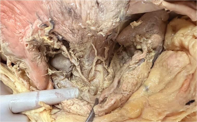
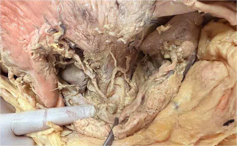
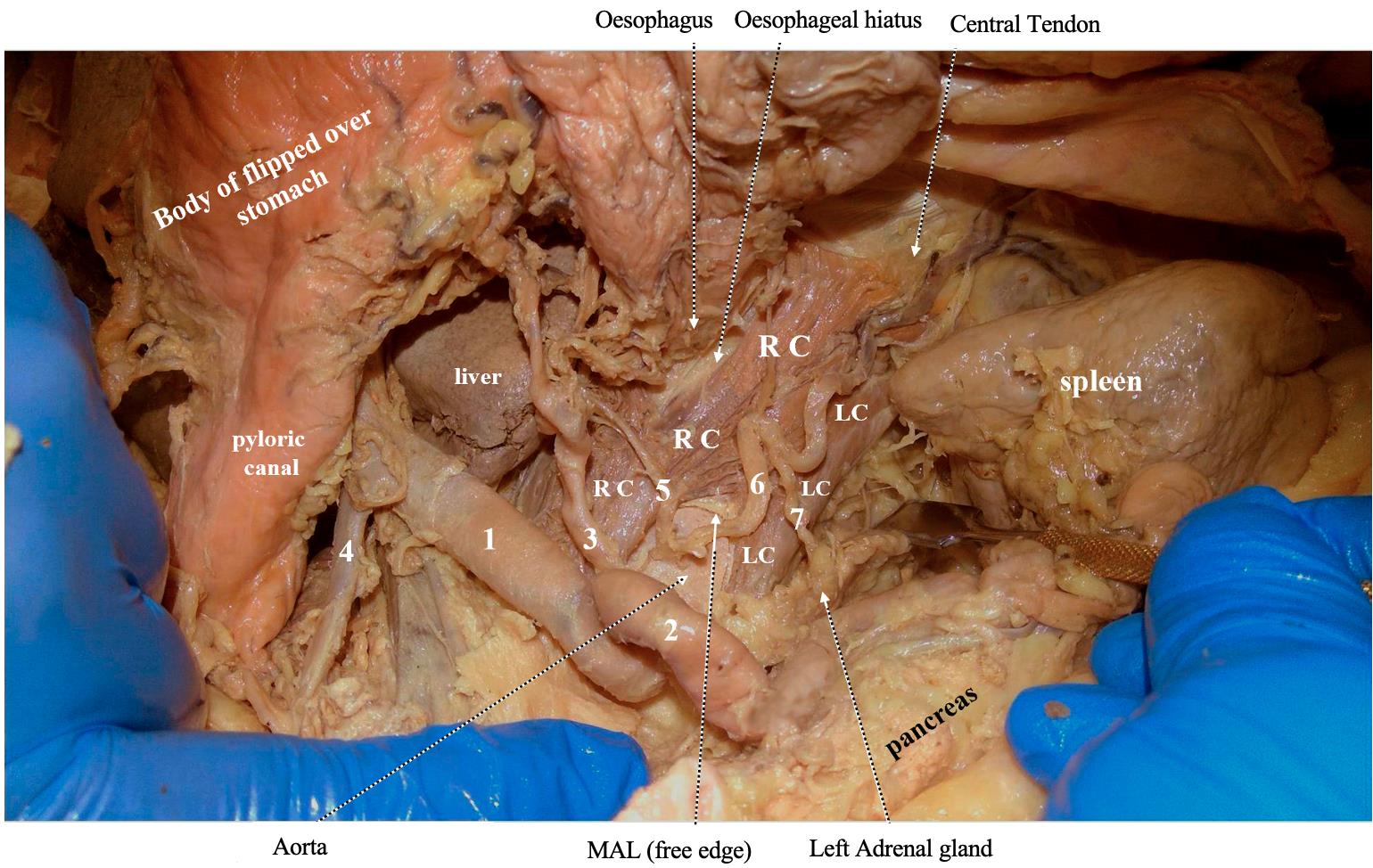
The median arcuate ligament (MAL) is at the core of a current controversy piqued by increased research queries regarding its contribution to the pathogenesis of coeliac artery compression syndrome. As our own curiosity was aroused by these queries, we decided to contribute to the discourse by providing a clear visual documentation of the MAL, obtained through cadaveric dissection in the Alfaisal University anatomy laboratory. We believe that reviewing the anatomy of the MAL, and the crura that form it, is crucial in this context. The cadaver was of a 50-year-old male and had been preserved in formalin. We used Grant’s Dissector Manual as a reference throughout the dissection, which was comprehensively documented in digital photographs as well as on video. Upon entering the lesser sac, we explored the stomach bed structures. We skeletonised the coeliac trunk up to the point of its emergence from the aorta. The peritoneum, the fascia covering the aorta, the wall, and the crura, as well as nearby sympathetic ganglia were carefully removed, resulting in the view seen in Figure 1. As much as we wished to leave the displayed anatomical features undisturbed per Figure 1, we also wanted to reveal the muscular and tendinous components of the diaphragmatic crura, to illustrate how their medial margins meet to form the MAL. Accordingly, some fascicles of the left crus were displaced backward, and a few of the right crus were removed (Figure 2). Normally, the highest end of the MAL wraps the anterior and lateral aspects of the aorta at the T12 vertebra, forming with the body of this vertebra the aortic hiatus (Figures 2 and 3). Due to MAL's wide and diverse placement, this ligament may lie low, compressing or distorting the coeliac trunk [1]. The coeliac trunk, also known as the ceoliac artery or truncus coeliacus, is a vessel arising
from the aorta. It passes beneath the MAL at the T12 vertebral level, which is also the point at which the aorta enters the abdominal cavity. Its three main branches are the left gastric artery, the common hepatic artery, and the splenic artery. The coeliac trunk provides a circulatory supply to the foregut, specifically the liver, pancreas, gall bladder, spleen, and distal oesophagus, up to the second portion of the duodenum [1]. Coeliac artery compression syndrome is commonly referred to as coeliac axis syndrome, median arcuate ligament syndrome (MALS), Dunbar syndrome, or Harjola-Marable syndrome. This distinctive condition arises from the compression of the coeliac artery by the median arcuate ligament [1]. Another factor that could contribute to the compression is the fibres of the coeliac ganglia [2]. Despite having been identified several decades ago, the existence of this syndrome is still disputed by several researchers. Harjola was the first to document the clinical importance of coeliac artery compression in a living person. In his 1963 paper, the author discusses the case of a 57- year-old man who experienced abdominal pain after eating. Further examination revealed that his symptoms were caused by the compression of the coeliac artery by a fibrotic coeliac ganglion, and the surgical excision of said ganglion led to the disappearance of the symptoms [3]. In 1967, Dunbar et al. released a series of cases in which comparable symptoms arose from external compression of the coeliac artery by fibres of the median arcuate ligament. Clinical improvement was observed after the surgical division of these fibres [4]. Patients with this condition typically present between the ages of 20 and 40, and it may also cause mesenteric ischaemia in certain people. Postprandial discomfort is the most prevalent complaint, in addition to nausea or vomiting and epigastric pain, which causes weight loss. Standing is usually the best way to reduce the pain, while lying down worsens it [1].
Ayman Behiery and Osama Ezzeldin Abdelhadi, are with the Department of Anatomy, Alfaisal University, Riyadh, Saudi Arabia, email: abehiery@alfaisal.edu, email: oabdelhadi@alfaisal.edu. Mohamad Bakir, Ahmad Dawalibi and M. Adnan AlDoumani are with the College of Medicine, Alfaisal University, Riyadh, Saudi Arabia, email: Mo7ammedbakir@gmail.com, email: ahmad.dawalibi@hotmail.com, email: maldoumani@alfaisal.edu. DOI: 10.52609/jmlph.v4i2.124
Intermittent mesenteric ischaemia is thought to be caused by the compression of the coeliac artery by the MAL. Nevertheless, this theory may not fully elucidate the situation, as an extensive collateral network of mesenteric arteries typically exists, connecting the coeliac artery to the superior mesenteric artery. Hence, malfunctioning of the coeliac nerve plexus could play a part in the development of this illness. Nerve failure can result in atypical constriction of the blood vessels supplying the abdominal organs, which can cause ischaemia [5]. Treatment for this illness frequently consists of laparoscopic surgical decompression, which is accomplished by separating the MAL, and is limited to symptomatic individuals [1]. Comprehending the complex connection between the MAL and the coeliac trunk is essential because of the latter’s critical function in systemic circulation. This artery, which arises from the aorta and is located below the MAL, supplies blood to vital abdominal organs. Thus, median arcuate ligament compression can have serious impacts on blood vessels and organ function. Our research highlights the limited availability on the internet of comprehensive anatomical illustrations of the MAL, despite the fact that precise anatomical photographs are crucial to clearly show the relationship between this ligament and the coeliac artery. Our focus involves a comprehensive cadaveric anatomical description to address this gap and to emphasise the significance of hands-on learning in anatomy instruction, promoting a more profound comprehension of the complexities related to the median arcuate ligament.
10.1016/j.avsg.2008.11.005. Epub 2009 Jan 6. PMID: 19128929.
3. Harjola PT. A rare obstruction of the coeliac artery. Report of a case. Ann Chir Gynaecol Fenn. 1963;52:547-50. PMID: 14083857. 4. Dunbar JD, Molnar W, Beman FF, Marable SA. Compression of the celiac trunk and abdominal angina. Am J Roentgenol Radium Ther Nucl Med. 1965 Nov;95(3):731-44. doi: 10.2214/ajr.95.3.731. PMID: 5844938. 5. Saleem T, Katta S, Baril DT. Celiac Artery Compression Syndrome. 2023 Apr 26. In: StatPearls [Internet]. Treasure Island (FL): StatPearls Publishing; 2024 Jan–. PMID: 29262206.
APPENDIX Access the video and visually explore these structures by following the link provided: [https://youtu.be/6hCzBl9w7XA]
REFERENCES 1. Ahluwalia N, Nassereddin A, Futterman B. Anatomy, Abdomen and Pelvis: Celiac Trunk. 2022 Sep 19. In: StatPearls [Internet]. Treasure Island (FL): StatPearls Publishing; 2023 Jan–. PMID: 29083567.
2. Duffy AJ, Panait L, Eisenberg D, Bell RL, Roberts KE, Sumpio B. Management of median arcuate ligament syndrome: a new paradigm. Ann Vasc Surg. 2009 Nov-Dec;23(6):778-84. doi: