Full text rendered from the published PDF. The PDF is the version of record; if the two differ, the PDF governs.
Negative pressure pulmonary oedema (NPPO) is a rare but serious complication for patients undergoing general anaesthesia. It often presents a diagnostic dilemma, thus posing significant management challenges. Despite the complexity of diagnosis, the pathophysiology and management of NPPO have been extensively studied. We present a case of NPPO in a middle-aged obese patient with high perioperative cardiac risk who underwent an emergency open appendicectomy. This case report details the anaesthetic approach to managing the patient’s complex comor-bidities, the series of events leading to the diagnosis, and subsequently the management of NPPO. We dis-cuss the challenges encountered in diagnosing and managing NPPO, and review potential preventive measures. Our conclusion underscores that diagnosis of NPPO remains challenging, and early identification of patients at risk with vigilant monitoring is crucial for timely diagnosis and effective management.
Keywords: Anaesthetics, Anaesthesia, General, Obesity, Pulmonary Oedema
Negative pressure pulmonary oedema (NPPO) is a rare but serious complication associated with gen- eral anaesthesia, with reported incidence ranging from 0.01% to 1% [1–4]. NPPO is characterised by noncardiogenic pulmonary oedema resulting from a sudden and significant reduction in intrathoracic pressure, and remains a diagnosis of exclusion. Despite its low incidence, NPPO often leads to life- threatening post-operative pulmonary complication s, Wing Hoh Loo (wing_hoh88@hotmail.com), Depart- ment of Anaesthesiology and Critical Care, Hospital Sungai Buloh; Kay Mint Goh (kaymint2003@ya- hoo.com), Department of Anaesthesiology and Critical Care, Hospital Ampang. DOI: 10.52609/jmlph.v5i2.175
particularly during the conclusion of anaesthesia [3]. We report a case of NPPO in a 59-year-old obese pa- tient with high perioperative cardiac risk, who un- derwent an emergency surgical procedure. Knowledge of such cases can increase awareness of the existence of NPPO, allowing for prompt man- agement and avoiding unnecessary treatment.
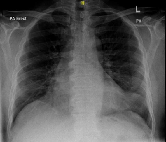
A 59-year-old gentleman was admitted to a tertiary centre with an acute abdomen. He presented with multiple comorbidities, including type II diabetes mellitus on insulin treatment, hypertension, obesity with body mass index (BMI) of 35, probable ob- structive sleep apnoea (OSA) indicated by a STOP BANG score of 5, and history of ischaemic heart dis- ease (IHD) with two vessels stented nine months ago. He was on dual antiplatelet therapy at the time of presentation. The STOP-BANG score, a widely used screening tool for OSA, is routinely applied in our practice, with a score greater than 4 suggesting a high probability of the condition [5]. A contrast-enhanced computed tomography (CT) scan revealed features indicative of acute appendici- tis, leading to the decision for an emergency open appendicectomy. Pre-anaesthetic assessment demonstrated a favourable cardiac status, with more than 4 metabolic equivalents (METS). A point-of- care echocardiogram revealed normal left ventricu- lar (LV) contractility and chamber sizes. Physical examination was largely unremarkable aside from obesity, with no sign of heart failure. Airway assess- ment indicated a Mallampati score of 1. The patient was stable haemodynamically, with normal satura- tion and normal chest X-ray (Figure 1). Based on the American Society of Anesthesiologists (ASA) phys- ical status classification, and considering his medi- cal history and comorbidities, he was classified as ASA 3E and scheduled for general anaesthesia with opioid sparing analgesia.
Modified rapid sequence induction (RSI) was em- ployed to induce anaesthesia. The patient was in- duced with intravenous (IV) lignocaine 100 mg, IV fentanyl 50 μg, IV propofol 110 mg, and IV rocu- ronium 100 mg. Intubation was assisted by video laryngoscope, with a percentage of glottis opening (POGO) score of 100%. Following induction, a uni- lateral right-sided transversus abdominis plane (TAP) block was performed under ultrasound guid- ance, administering 20 ml levobupivacaine 0.25%. Anaesthesia was maintained with sevoflurane, with O2/N2O titrated to achieve an age-adjusted minimal alveolar concentration (MAC) of 0.8. As part of his analgesic regimen, the patient received IV dexame- thasone 8 mg, IV magnesium sulphate 20 mmol, IV oxycodone 2 mg, and IV paracetamol 1 g upon sur- gical incision. Intraoperatively, he was diagnosed with a perforated appendix without abdominal con- tamination. The patient received a total of 800 ml crystalloid over the course of the two-hour surgery. Notably, no additional rocuronium top-up was re- quired throughout the procedure. Upon conclusion of the surgery, the patient was re- versed with IV neostigmine 2.5 mg and IV glyco- pyrrolate 400 μg upon return of spontaneous regular breathing. Prior to extubation, the patient exhibited responsiveness to commands, and maintained ade- quate saturation with satisfactory spontaneous ven- tilation. Additionally, there was evidence of robust power recovery clinically, exemplified by sustained head lift and hand grip for longer than 5 seconds. However, upon extubation, he experienced shortness of breath and desaturated to 93%. At that point, his blood pressure (BP) measured 128/72 mmHg, heart rate (HR) was 75 beats per minute (bpm), and aus- cultation of his lungs revealed bilateral basal coarse crepitations. Immediate intervention included ad- ministration of IV frusemide 5 mg and the applica- tion of continuous positive pressure (CPAP) at 5 cmH20 with 100% O2. Subsequently, his saturation improved, and he was monitored in the Post Anaes- thetic Care Unit (PACU). In the PACU, while on a face mask at 5 L/min O2, the patient had another episode of desaturation with
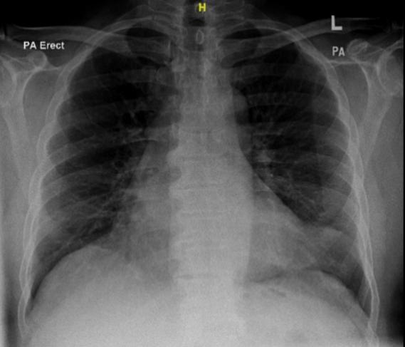
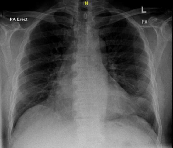
SPO2 dropping to 92%, with evidence of a shunt. Examination of the lungs revealed coarse crepita- tions over bilateral lower zones. His BP was 140/85, HR was 82 bpm, and arterial blood gas (ABG) showed type 1 respiratory failure with lactic acid- osis. He received another bolus of IV frusemide 10 mg, and non-invasive ventilation (NIV) was initi- ated. An urgent portable chest X-ray was ordered, revealing features consistent with pulmonary oe- dema (Figure 2). Consequently, the patient was admitted to the Inten- sive Care Unit (ICU) with a working diagnosis of unstable angina complicated with acute pulmonary oedema. In the ICU, his condition gradually im- proved on NIV, and on the same day of admission, he was successfully weaned to nasal prong O2 sup- plementation. Repeated chest X-ray revealed a pic- ture of resolving pulmonary oedema (Figure 3). Se- rial bedside echocardiography and ECG showed no signs of acute myocardial ischaemia, and the pa- tient’s serial troponin I level remained static — not suggesting a cardiac aetiology of acute pulmonary oedema. Ultimately, the patient was discharged from ICU on the second day of admission, and discharged from the hospital in stable condition on the fifth day of hospitalisation.
NPPO is an acute life-threatening perioperative complication. While establishing a definitive diag- nosis may not immediately alter management, it plays a crucial role in avoiding harmful interven- tions, such as the inappropriate use of antiplatelet therapy in cases misdiagnosed as acute coronary syndrome (ACS). A precise diagnosis also ensures timely and targeted treatment, improving patient outcomes.
The classical description of the pathophysiology of NPPO involves the occurrence of acute pulmonary oedema following excessive negative intrathoracic pressure as a result of deep inspiration over an ob- structed upper airway; in most cases, over a closed glottis [3,6]. Although the reported incidence of
NPPO is low, it is likely underreported due to the difficulty in confirming the diagnosis, and to its typ- ically rapid, self-resolving clinical course [7]. The aetiology of NPPO can be broadly classified into acute and chronic airway obstruction. Conditions leading to acute upper airway obstruction, such as
Two mechanisms have been proposed to explain the pathogenesis of NPPO [3,7]. Oswalt and colleagues (1977) proposed that the sudden fluctuations in in- trathoracic pressure result in significant fluid shifts [8]. This is described as a combination of increased preload and afterload, in addition to a decrease in pulmonary interstitial pressure, ultimately leading to a substantially high hydrostatic pulmonary pressure gradient. According to Starling forces, this results in transudation of fluid into the interstitium from the pulmonary capillary [6]. West and colleagues (1992) described the concept of wall stress failure more than 30 years ago [9]. The increase in transmural pulmonary capillary pressure leads to disruption of the alveolar-capillary membrane, increasing the per- meability and resulting in high-protein pulmonary oedema. It is important to understand the possible pathogenesis to ensure a targeted management plan. In this case, a cardiogenic cause of acute pulmonary oedema (APO) was the primary differential diagno- sis, in view of the patient’s high revised cardiac risk index (RCRI). This was subsequently ruled out, based on serial biochemical markers and ECG find- ings. Given the risks associated with initiating an- tiplatelet therapy immediately post-operatively, and
post-extubation laryngospasm, croup, and epiglotti- tis, can predispose individuals to NPPO. On the other hand, chronic upper airway obstructions re- sulting from obesity, obstructive sleep apnoea, and any anatomical pathologies, may increase the risk of developing NPPO.
observing that the patient's condition improved sig- nificantly with NIV support, the decision was made to withhold antiplatelets unless ACS was defini- tively confirmed. However, statin therapy was promptly resumed in line with standard recommen- dations. Another differential considered was inadequate re- versal of the neuromuscular blockade. Although the patient demonstrated clinically adequate recovery prior to extubation, perioperative neuromuscular function was not quantitatively monitored, either in- traoperatively or during the event. Clinical assess- ments alone may be inadequate, as evidence sug- gests that a substantial proportion of patients who seem clinically recovered do not achieve a train-of- four ratio (TOFR) >90%, which poses a significant risk for unsafe extubation [10]. Consequently, we cannot completely exclude residual paralysis or re- curarisation in this case. The use of specific reversal agents, such as sugammadex, also warrants discus- sion, as there have been isolated reports of NPPO following its administration [11]. This underscores the importance of perioperative neuromuscular monitori ng, especially in high-risk patients, to mini- mise avoidable complications.
Other potential differential diagnoses which were considered include tracheal aspiration and anaphy- laxis. In this case, tracheal aspiration was unlikely as the patient was adequately fasted and was extubated awake at the end of anaesthesia. Similarly, the ab- sence of cutaneous allergic rash, mucosal angi- oedema, rale, or haemodynamic instability excluded the possibility of anaphylaxis [3,4]. In this patient, several clinical findings supported the diagnosis of NPPO, including respiratory distress at extubation, significant hypoxia, coarse crepitations on auscultation, and radiographic evidence of pul- monary oedema on chest X-ray [7]. The patient’s un- derlying obesity and possible history of obstructive sleep apnoea further increased the likelihood of NPPO. Negative findings, such as normal sequential troponin levels and the absence of regional wall mo- tion abnormalities on bedside echocardiography, helped to exclude cardiogenic causes. Ultimately, NPPO remains a diagnosis of exclusion. It is crucial to systematically rule out other differen- tials requiring immediate specific management, such as myocardial infarction, while avoiding inap- propriate and potentially harmful treatments. Careful precautions were taken to minimise the peri- operative cardiac and pulmonary risk in our patient, who was identified as high risk due to obesity with possible OSA and a high RCRI. Multimodal analge- sia including IV lignocaine, IV opioids, IV dexame- thasone, IV paracetamol, inhalational nitrous oxide, and regional anaesthesia, was employed to blunt sympathetic reflexes during intubation and surgical stimulation. These measures, alongside careful in- traoperative haemodynamic management and a con- trolled extubation process, aimed to reduce myocar- dial oxygen demand while maintaining adequate coronary perfusion. Fluid administration was care- fully monitored to avoid overload, and multimodal analgesia, supplemented by a regional block tech- nique, ensured optimal pain control. Additionally, muscle relaxant recovery was assessed clinically prior to extubation, although quantitative neuromus-
cular monitoring was not performed. These strate- gies were designed to optimise the balance between myocardial oxygen supply and demand.
However, the question remains: could anything have been done differently to prevent this incident? One key area for improvement is the use of neuromuscu- lar monitoring to objectively assess the adequacy of muscle relaxant recovery, especially given the un- predictable pharmacokinetics in obese patients. Quantitative monitoring could have provided a more reliable assessment, reducing the risk of inadequate recovery or residual neuromuscular blockade.
Several authors have suggested techniques to pre- vent NPPO. The strategies described include use of IV dexamethasone, IV lignocaine, and low-dose IV propofol prior to extubation [1,3,7]. The lesson from this case is early identification of risk factors and vigilant monitoring perioperative to facilitate prompt and effective management.
The use of sugammadex in patients with high RCRI has been advocated as it allows avoidance of anti- cholinergic agents which would undoubtedly cause tachycardia [12]. However, some case reports have noted NPPO following reversal with sugammadex [11]. It was postulated that there is a difference in the reversal time of upper airway muscles and the dia- phragm. Therefore, NPPO develops when there is rapid recovery of respiratory forces in the presence of upper airway collapsibility [11]. This highlights the complexity of managing high-risk patients and the need for tailored approaches to optimise out- comes while mitigating risks.
Management
The general management of NPPO is similar to that of cardiogenic pulmonary oedema, targeted to cor- rect hypoxia, with addition measures to correct up- per airway obstruction [1]. The specific manage- ment of NPPO includes the application of CPAP by means of NIV or, in rare cases, reintubation [1,6,13,14]. CPAP will reverse the fluid shift by re- ducing the fluctuation in transmural pulmonary
capillary pressure. The use of loop diuretics, specifically frusemide, has been widely reported in case reports and case series on NPPO. Despite its common use, unlike in cardi- ogenic pulmonary oedema, evidence of its benefit in NPPO is scarce [3,7]. Our argument for its use would be its effect in reducing preload and afterload, which would then reduce the hydrostatic pulmonary pressure gradient [15]. In this case, CPAP was applied via non-invasive ventilation (NIV), and titrated boluses of frusemide were administered. The patient showed rapid clini- cal improvement while further investigations were carried out to establish a definitive diagnosis. An- tiplatelet therapy was deliberately withheld after carefully weighing the benefits against the risks of bleeding, a decision ultimately justified as acute cor- onary syndrome (ACS) was ruled out. Overall, the management approach was appropriate for immedi- ate intervention and could serve as a reference for developing a structured algorithm for perioperative APO.
Diagnosing NPPO remains a challenge clinically, especially in patients with multiple comorbidities. The aetiology and pathophysiology have been well described, allowing for early identification of pa- tients at risk. We advocate vigilant perioperative monitoring in such patients, with mandatory neuro- muscular monitoring where available. With early di- agnosis, targeted management of airway reestablish- ment and oxygenation by positive airway pressure are essential to prevent complications while avoid- ing unnecessary interventions that could potentially cause harm.