Full text rendered from the published PDF. The PDF is the version of record; if the two differ, the PDF governs.
A 73-year-old man on chronic warfarin therapy presented to the emergency department with acute right thigh pain and swelling, without a history of trauma. Musculoskeletal point-of-care ultrasound (MSK-POCUS) rapidly identified an intramuscular hematoma with evidence of active hemorrhage. Subsequent computed tomography angiography (CTA) confirmed a sizable hematoma in the right vastus intermedius muscle and detected a small pseudoaneurysm. The patient was managed conservatively, with close monitoring yielding no further complications. This case demonstrates the diagnostic utility and efficiency of MSK-POCUS in the acute evaluation of musculoskeletal complaints, particularly in patients at risk for anticoagulant-induced bleeding. The report highlights the value of bedside ultrasound for prompt diagnosis, its advantages over other imaging modalities in emergency settings, and the importance of enhancing training for widespread POCUS implementation in clinical practice.
Keywords: Anticoagulants, Hematoma, Point-of-Care, Thigh, Ultrasonography, Warfarin
Acute haemorrhage is a major complication of oral anticoagulant (OAC) therapy, and could be so se- vere as to threaten the patient's life. Management, including reversal of the OAC, depends on the ur- gency, site, and severity of the haemorrhage. This case report emphasises the significance of integrat- ing musculoskeletal point-of-care ultrasound (MSK-POCUS) into the initial assessment of mus- culoskeletal complaints in the acute setting, to de- tect the presence of intramuscular haemorrhage. Ahlam Alfahmi (alfahmi.a.m@gmail.com) and Shahad Sheerah (Sasheerah@kfmc.med.sa) are with the Emergency Medicine Department, King Fahd Medical City, Riyadh, Saudi Arabia. DOI: 10.52609/jmlph.v5i4.190
Ahlam Alfahmi, Shahad Sheera
History of Presenting Illness A 73-year-old male presented to the emergency de- partment (ED) complaining of a sudden onset of sharp pain in the right thigh that started two days prior while he was sleeping. The course of the pain was progressive. The patient denied any history of trivial trauma, loss of consciousness, recent travel, immobilisation, fever, skin changes, chest pain, shortness of breath, palpitations, or previous simi- lar history.
Past Medical Illness The patient was known to have valvular heart dis- ease, post-mitral valve replacement, and atrial flut- ter. He was on warfarin and bisoprolol, and his INR fluctuated between 2.2 to 3. The last INR before the current ED visit was 4.2, and his warfarin dose was decreased from 5 mg to 4.5 mg.
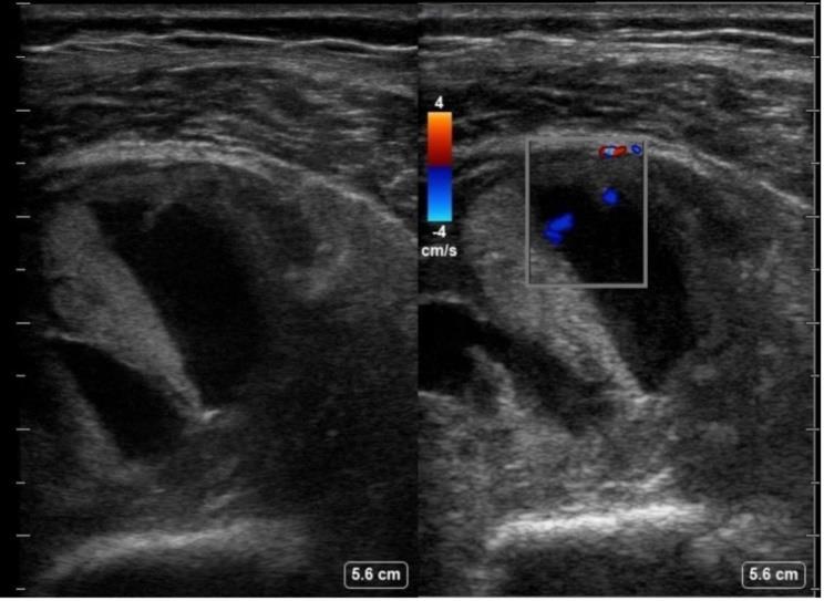
Physical Examination The patient was conscious, alert, oriented, looked well, and was calm in the bed. His blood pressure was 132/88 mmHg, heart rate 72 bpm, respiratory rate 20 bpm, and temperature was 37 °C. Physical examination of the right lower limb showed that the right thigh was slightly swollen by visual estima- tion compared with the left thigh, with no skin col- our changes or hotness, and the compartments were firm and tender. Neurovascular examination was unremarkable. Using a linear probe (6-13 mHz) and on musculo- skeletal setting, the ultrasound ( Supplementary Video 1a and 1b ) illustrated a well-defined hy- poechoic area within the lateral aspect of the upper part of the thigh, which suggests a collection. Within the collection, an internal flow is noted when colour Doppler was applied, suggesting an active haemorrhage ( Supplementary Video 2 and figure 1).
Laboratory Examinations The complete blood count (CBC) results were as follows: haemoglobin 9.3 g/dL (no significant drop compared with a previous result of 9.6 g/dL);
haematocrit 29%; mean corpuscular volume (MCV) 97.3 pg; mean corpuscular haemoglobin (MCH) 31.2 g/dL. The coagulation profile revealed a partial thromboplastin time (PTT) of 41 seconds, prothrombin time (PT) of 35.1 seconds, and inter- national normalised ratio (INR) of 2.73. The renal profile showed a creatinine level of 329 μmol/L, urea 24.9 mg/dL, and potassium 6.3 mEq/L.
Imaging Examination CTA of the lower extremity showed a 3 x 6.3 x 9.3 cm heterogeneous collection in the right vastus in- termedius muscle, with a small, 0.4 cm pseudoan- eurysm in the anterior aspect of the lesion, likely arising from the terminal branches of the profunda femoris artery.
Treatment Course This case involved a warfarin-associated intramus- cular hematoma with active bleeding from a branch of the profunda femoris artery. It was managed conservatively using a watch-and-wait approach; after 11 days, there were no further complications or progression of the hematoma. The patient's hae- moglobin remained stable, and his INR was within the therapeutic range. Because he was at high risk for thrombosis, the primary team resumed warfa- rin. The patient was discharged in stable condition with regular follow-up scheduled in the outpatient department.
We report a case of spontaneous muscle hematoma diagnosed at the bedside using MSK-POCUS. Such hematomas are most commonly associated with an- ticoagulant use and have a higher prevalence among elderly patients [1]. While imaging modali- ties such as CT and MRI are often utilised for diag- nosis [2], ultrasound offers a rapid, efficient, safe, and accessible alternative that can complement the clinical examination. In cases of acute bleeding, ul- trasound allows for the objective identification of intramuscular blood collections, accurate measure- ment of their size and location, monitoring of their progression, and confirmation of resolution [3]. Typical ultrasound findings include disruption of the muscle’s normal fibrillar pattern, with anechoic and hypoechoic areas indicating the presence of blood [3]. MSK-POCUS is specifically designed for the tar- geted assessment of musculoskeletal structures, utilising limited scanning planes or simplified pro- tocols to streamline the evaluation process [4]. It is
frequently integrated into the ED workflow as an extension of the physical examination, providing dynamic, high-resolution imaging of muscles, soft tissues, and joints through high-frequency trans- ducers [3.5–15 MHz] [5]. Although MRI is considered the gold standard for diagnosing soft tissue conditions [6], its routine use is limited by factors such as restricted availability, lengthy examination times, the need for sedation in paediatric patients, and significant cost [2]. These limitations make ultrasound—particularly MSK- POCUS—a more practical and prevalent option in many acute care settings. The clinical value of MSK-POCUS is further sup- ported by the case reported by Sharma et al. [7], in which an iliopsoas haemorrhage was identified in an elderly patient on warfarin, using POCUS in the ED. This exemplifies the utility of MSK-POCUS in promptly identifying deep intramuscular haem- orrhages. Recent advancements in bedside imaging include the use of contrast-enhanced ultrasound (CEUS) [8]. For instance, Ma et al. [9] demonstrated the ef- fectiveness of bedside CEUS in a patient with left upper limb pain and swelling, where it revealed a hematoma with active haemorrhage from the bra- chial artery. CEUS has proved to be a rapid and ac- curate tool for detecting microvascular and low-ve- locity haemorrhages, and holds promise as an alter- native imaging modality when CT is contraindi- cated or unavailable. IV. In our case, MSK-POCUS enabled the rapid as- sessment of the patient’s symptoms and the detec- tion of active haemorrhage, which was subse- quently confirmed with additional imaging. Never- theless, limitations are expected due to the nature of the case report study. Initial MSK-POCUS com- pression and follow-up scan could have provided valuable information regarding the hematoma’s progression or resolution. Additionally, a key area for improvement was the integration of MSK-PO- CUS into emergency training programs to enhance the effective use of this diagnostic tool.
In conclusion, incorporating POCUS into routine physical examinations including MSK-POCUS can significantly enhance the detection of soft tissue pathologies, potentially shortening ED stays and enabling more rapid clinical decision-making and timely interventions. However, POCUS remains
highly operator-dependent. Integrating POCUS into training programs can help standardise prac- tice among operators, promoting more consistent and reliable use of the modality.
V. REFERENCES 1. Dohan A, Darnige L, Sapoval M, Pellerin O. Spontaneous soft tissue hematomas. Diag- nostic and Interventional Imaging . 2015 Jul;96(7-8):789-96. 2. Querol F, Rodriguez-Merchan EC. The role of ultrasonography in the diagnosis of the musculoskeletal problems of haemophilia. Haemophilia : the official journal of the World Federation of Hemophilia . 2012 May;18(3):e215-26. 3. de la Corte-Rodriguez H, Rodriguez-Mer- chan EC. Treatment of muscle haematomas in haemophiliacs with special emphasis on percutaneous drainage. Blood Coagulation & Fibrinolysis . 2014 Dec;25(8):787–94. 4. Bakeer N, Shapiro AD. Merging into the mainstream: the evolution of the role of point-of-care musculoskeletal ultrasound in hemophilia. F1000Research . 2019;8. 5. Bailey C, Bakeer N. Practice guidelines for implementation and use of musculoskeletal ultrasound in the management of individuals with hemophilia and other bleeding disor- ders. J Clin Orthop Trauma . 2020;11(4):645- 654. doi:10.1016/j.jcot.2020.06.016. 6. Mayerson JL, Scharschmidt TJ, Lewis VO, Morris CD. Diagnosis and management of soft-tissue masses. Instructional Course Lec- tures. 2015;64:95-103. 7. Sharma D, Saker R, Govind A. Emergency department ultrasound diagnosis of sponta- neous iliopsoas hemorrhage in a patient on warfarin. BMJ Case Reports. 2013 Nov 15;2013. 8. Miele V, Piccolo CL, Galluzzo M, Ianniello S, Sessa B, Trinci M. Contrast enhanced ul- trasound (CEUS) in blunt abdominal trauma. The British Journal of Radiology . 2016;89(1061):20150823. 9. Ma JJ, Zhang B. Diagnosis of an actively bleeding brachial artery hematoma by con- trast-enhanced ultrasound: A case report.
World Journal of Clinical Cases. 2020 Sep 6;8(17):3835-40.
Supplementary Material Supplementary Video 1a and b : B-mode ultra- sound view shows a well-defined hypoechoic area within the muscle, consistent with an intra- muscular collection with an internal flow. Supplementary Video 2 : Colour Doppler imag- ing reveals a colour signal within the hematoma suggestive of active internal bleeding.