Full text rendered from the published PDF. The PDF is the version of record; if the two differ, the PDF governs.
Prompt diagnosis and treatment of neonatal septic arthritis (SA) is crucial to prevent severe complications such as joint deformity and long-term disability. This report details the case of a 16-day-old male neonate presenting with localised swelling, erythema, and restricted movement of the right knee, ultimately diagnosed with Escherichia coli -induced septic arthritis—a previously undocumented finding in Saudi Arabia. The diagnosis was confirmed via synovial fluid culture, and management included the administration of broad-spectrum antibiotics adjusted according to sensitivity, as well as surgical intervention. This case highlights the significance of an early, multidisciplinary approach in managing neonatal septic arthritis and adds to the literature on atypical causative pathogens.
Keywords: Arthritis, Infectious, Neonate, Pediatric Emergency, Saudia Arabia
Neonatal septic arthritis (SA) presents unique challenges in diagnosis and treatment, especially in preterm infants who are at higher risk due to immature immune systems and hospital expo- sures. While most cases are caused by pathogens such as Staphylococcus aureus and Klebsiella pneumoniae , this report documents a rare instance of SA caused by Escherichia coli , the first such case reported in Saudi Arabia. This underscores the importance of recognising risk factors and em- ploying a multidisciplinary management ap- proach.
A 16-day-old boy presented to the emergency Khalid AlMahmoud (K.almahmoud21@gmail.com) is with the Paediatric Department, King Saud University Medical City, Riyadh, Saudi Arabia. Lama Alfakhri (lama.alfakhri@gmail.com); Mashel A. Hussain (Mashael.A.K.H@gmail.com); and Amal Yousif (dr.amalyousif@gmail.com) are with the Paediatric Depart- ment, King Saud University Medical City, Riyadh, Saudi Arabia. Abdulrahman Alzahrani (aakz1415@gmail.com) is with the Paediatric Department of Emergency Medicine, Emergency Administration, King Fahd Medical City, Sec- ond Health Cluster, Riyadh, Saudi Arabia. DOI: 10.52609/jmlph.v5i4.193
department (ED) with localised swelling, ery- thema, warmth, and limited motion of the right knee that had continued for one day. There was no history of trauma, fever, or systemic symptoms. The patient was born prematurely at 34 weeks via emergency caesarean section (C-Section) due to foetal distress following the mother's preterm premature rupture of membranes (PPROM), which was managed appropriately. Post-delivery, he was admitted to the neonatal intensive care unit (NICU) for management of respiratory distress and low birth weight (1.8 kg). He was discharged after seven days and scheduled for regular follow- up visits at the neonatal clinic to monitor growth, development, and overall health. Upon presentation to the ED, the patient appeared active and was not in generalized pain or distress. However, he became irritable when his right leg was manipulated, indicating localized discomfort. He was neither pale, cyanosed, not jaundiced. Ac- cording to the parents, the infant had a single epi- sode of mild fever (approximately 38.0°C) the day before presentation, which resolved sponta- neously and was not present upon arrival at the ED. His vital signs were as follows: rectal temper- ature 37.7°C, respiratory rate 38 breaths per mi- nute, SpO 2 98%, and body weight 2.2 kg. The fon- tanelle was normal in size, and the suture lines were palpable. Tearing of the eyes and moist mu- cous membranes were observed. The extremities were warm, and peripheral pulses were palpable bilaterally. Respiratory, abdominal, and cardio- vascular examinations were unremarkable. Geni- tal examination revealed an uncircumcised penis with hypospadias. No lymphadenopathy, ulcers, or rashes were observed. The neonate presented with significant swelling and erythema of the right knee, accompanied by a restricted range of motion. Despite these findings, vital signs were within normal range, and the sys- temic examination revealed no other abnormali- ties. The emergency team initiated further evalu- ation with laboratory tests and imaging, and con- sulted the orthopaedics team, considering septic arthritis versus osteomyelitis as the leading differ- ential diagnoses.
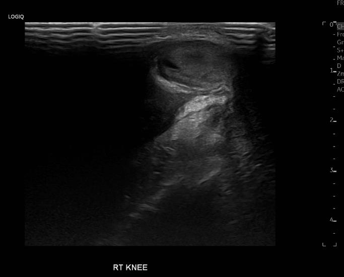
Blood tests revealed an elevated C-reactive pro- tein (CRP) level of 99.68 mg/L and erythrocyte sedimentation rate (ESR) of 24 mm/hr, both sug- gesting an inflammatory response. A knee ultrasound (Figure 1) revealed joint effu- sion and surrounding soft tissue hyperaemia, find- ings consistent with SA. Empirical antibiotic ther- apy was initiated with cefotaxime (50 mg/kg every 8 hours) and ampicillin (100 mg/kg every 12 hours), and was continued for three days pend- ing the results of diagnostic investigations. Joint aspiration, performed in the operating room by the orthopaedics team, revealed purulent fluid. Synovial fluid cultures subsequently identified Escherichia coli as the causative pathogen. Ac- cordingly, antibiotic therapy was adjusted based on culture sensitivity to cefepime (50 mg/kg every 12 hours) and vancomycin (15 mg/kg every 8 hours), which were administered for a total dura- tion of 10 days. The patient tolerated all medica- tions and procedures well, with no reported ad- verse effects or complications throughout his hos- pitalisation. At the 3-month follow-up visit, the infant demonstrated normal growth parameters, full range of motion of the affected joint, and no signs of recurrent infection or developmental de- lay. This highlights the importance of a multidis- ciplinary approach in managing complex cases of neonatal septic arthritis.
hyperaemia, findings consistent with septic arthritis
Neonatal septic arthritis is a rare but critical diag- nosis in the neonatal period, typically resulting from a haematogenous spread of infection. De- layed recognition can lead to irreversible joint damage, including growth disturbances, avascular necrosis, and long-term functional impairment. Early diagnosis and targeted antimicrobial ther- apy, along with timely surgical intervention when indicated, are essential to optimise outcomes [1,2]. Previous studies have emphasised that neonatal SA typically manifests with vague, nonspecific symptoms, such as irritability, feeding difficul- ties, and restricted limb movement, which can complicate clinical diagnosis. This aligns with our case, where localized irritability upon manipula- tion and limited mobility of the affected limb were the main presenting features. Although the parents reported a brief, intermittent fever epi- sode, it was absent during clinical evaluation, and other classic signs of infection were also lacking [3,4]. Neonatal SA is strongly associated with risk fac- tors such as prematurity and invasive procedures, including C-section and umbilical catheterisation. In this case, the patient was a 34-week preterm in- fant delivered via C-section who required NICU admission, placing him at heightened risk for SA.
Another study further highlights that preterm ne- onates are more prone to SA, likely owing to their immature immune systems and higher exposure to hospital-acquired infections [5]. The gold standard for diagnosing the condition is a synovial fluid culture [6]. A study of neonatal SA found that Staphylococcus aureus was the most frequently isolated pathogen, followed by Klebsiella pneumoniae and Klebsiella oxytoca [7]. However, the microbio- logical findings in this case identified E. coli in the synovial fluid culture, which is rare and not previously documented in the literature. To the best of our knowledge, this is the first case from Saudi Arabia to report neonatal SA with E. coli as the causative pathogen. The management of neonatal SA typically in- volves initiating empirical antibiotic therapy to target the most likely pathogens, followed by cul- ture-guided adjustments once the causative organ- ism is identified. In this case, the neonate was in- itially treated with a broad-spectrum regimen of cefotaxime and ampicillin consistent with current guidelines for SA management [8]. After identi- fying E. coli as the causative agent, the antibiotics were adjusted accordingly to cefepime and van- comycin. This strategy aligns with recommendations from other studies that advocate initiating broad-spec- trum antibiotics in suspected cases of neonatal SA. While antibiotic therapy is crucial, early sur- gical intervention is often necessary to address joint effusion and mitigate further complications. In our case, the neonate underwent arthrotomy for joint drainage and debridement, a common proce- dure for managing SA with significant joint effu- sion. Evidence suggests that combining timely surgical intervention with appropriate antibiotic therapy significantly reduces the risk of long-term complications, such as joint deformities and chronic infection [7]. This case underscores the importance of early de- tection and management in cases of neonatal SA, especially in preterm infants at elevated risk. The identification of E. coli as the causative pathogen broadens the spectrum of organisms linked to this condition. Effective management, including timely diagnosis, appropriate antibiotic therapy, and surgical intervention, is essential to prevent long-term complications. In our case, multidisci- plinary collaboration was key to the successful
outcome, emphasising vigilance in identifying uncommon pathogens in similar cases. This case report is limited by its nature as a single- patient observation, which restricts the generali- sability of findings. Although E. coli was identi- fied as the causative organism, broader conclu- sions about its prevalence in neonatal SA cannot be drawn. Additionally, the absence of long-term follow-up data limits assessment of functional outcomes and potential joint sequelae. Further multicentre studies are needed to better character- ise atypical pathogens and optimal management strategies in neonatal septic arthritis.
Ethical Considerations Ethical approval was obtained from the Institu- tional Review Board of King Fahad Medical City, Riyadh Second Health Cluster, Saudi Arabia (IRB Registration Number: IRB00010471). Written in- formed consent for publication of the case details and images was obtained from the patient’s legal guardian, in accordance with the Declaration of Helsinki and institutional guidelines.
IV. FUNDING SOURCES None to declare.
V. REFERENCES 1. Almatrafi MN, Almatrafi MA, Qronfla HM, Osaylan MT, Bajuifer SA. Neonatal septic arthritis of the hip: A case report. Cu- reus. 2023 Jul 31;15(7):e42738. doi: 10.7759/cureus.42738. PMID: 37654918; PMCID: PMC10467518. 2. Rai A, Chakladar D, Bhowmik S, Mondal T, Nandy A, Maji B, Hazra A, Mondal R. Neonatal septic arthritis: Indian perspec- tive. Eur J Rheumatol. 2020 Feb;7(Suppl1):S72-S77. doi: 10.5152/eur- jrheum.2019.19052. PMID: 35929862; PMCID: PMC7004267. 3. Liu Y, Zhao K, Liu Y, Sun YH, Li MX, Yu M, Zhu LQ, Wang XD. Bone and joint in- fection complicated with sepsis in neonates and infants under three months of age. J Pe- diatr (Rio J) . 2024 Mar-Apr;100(2):156- 162. doi: 10.1016/j.jped.2023.09.003. Epub 2023 Oct 12. PMID: 37837994; PMCID: PMC10943287. 4. Gatto A, Lazzareschi I, Onesimo R, Ian- notta R, Rigante D, Capossela L, Filoni S, Valentini P. Short therapy in a septic arthri- tis of the neonatal hip. Pediatr Rep . 2019
Sep 27;11(3):8161. doi: 10.4081/pr.2019.8161. PMID: 31588259; PMCID: PMC6775484. 5. Sukswai P, Kovitvanitcha D, Thumkunanon V, Chotpitayasunondh T, Sangtawesin V, Jeerathanyasakun Y. Acute hematogenous osteomyelitis and septic ar- thritis in children: clinical characteristics and outcomes study. J Med Assoc Thai. 2011 Aug;94 Suppl 3:S209-16. PMID: 22043778. 6. Pammi M, Flores A, Versalovic J, Leeflang MM. Molecular assays for the diagnosis of sepsis in neonates. Cochrane Database Syst Rev . 2017 Feb 25;2(2):CD011926. doi: 10.1002/14651858.CD011926.pub2. PMID: 28236648; PMCID: PMC6464551. 7. Li Y, Zhou Q, Liu Y, Chen W, Li J, Yuan Z, Yong B, Xu H. Delayed treatment of sep- tic arthritis in the neonate: A review of 52 cases. Medicine (Baltimore) . 2016 Dec;95(51):e5682. doi: 10.1097/MD.0000000000005682. PMID: 28002339; PMCID: PMC5181823. 8. Sreenivas T, Nataraj AR, Kumar A, Menon J. Neonatal septic arthritis in a tertiary care hospital: a descriptive study. Eur J Orthop Surg Traumatol . 2016 Jul;26(5):477-81. doi: 10.1007/s00590-016-1776-9. Epub 2016 May 6. PMID: 27154290.