Full text rendered from the published PDF. The PDF is the version of record; if the two differ, the PDF governs.
Background: Button battery ingestion is a paediatric emergency associated with significant morbidity and mortality. Rapid recognition and removal are critical to prevent severe oesophageal and mediastinal complications.
Methods: We report a case series of five patients with a mean age of 22.6 months (SD= 11.4 months) with confirmed button battery ingestion who were managed at a tertiary referral center.
Results: Patients ranged from 13 to 41 months of age. Presentations varied from asymptomatic delayed presentation to acute respiratory distress. Endoscopic removal was required in all cases, with findings ranging from superficial mucosal erythema to severe grade 3 caustic necrosis. One child demonstrated features suggestive of an impending aortoesophageal fistula. Supportive care included parenteral nutrition, antibiotics, and proton pump inhibitors. Despite successful retrieval in most of the cases, long-term complications remain a concern.
Conclusion: Button battery ingestion in children is unpredictable both in presentation and in outcome. Delays in recognition and failed initial removal attempts are associated with higher grades of oesophagal injury. Based on these findings, we propose the establishment of a “Button Battery Code” to standardise rapid response, expedite referral, and coordinate multidisciplinary management, similar to STEMI and stroke codes.
Keywords: Button Battery, Burns, Chemical, Child Preschool, Emergency Treatment, Endoscopy, Esophageal Injuries, Foreign Bodies
Button battery ingestion is a pediatric emergency associated with significant morbidity and mortality. Rapid recognition and removal are critical to prevent severe esopha- geal and mediastinal complications. Methods: We report a case series of five patients with a mean age of 22 months (SD= 11.4 months) with confirmed button battery ingestion who were managed at a tertiary referral center. Results: Patients ranged from 13 to 41 months of age. Presentations varied from asymptomatic delayed presentation to acute respiratory distress. Endoscopic removal was required in all cases, with findings ranging from superficial mucosal erythema to severe grade 3 caustic necrosis. One child demonstrated features suggestive of an impending aortoesophageal fistula. Support- ive care included parenteral nutrition, antibiotics, and proton pump inhibitors. Despite success- ful retrieval in most of the cases, long-term complications remain a concern. Conclusion: Button battery ingestion in children is unpredictable both in presentation and in outcome. Delays in recognition and failed initial removal attempts are associated with higher grades of esophageal injury. Based on these findings, we propose the establishment of a “Button Battery Code” to standardize rapid response, expedite referral, and coordinate multidisciplinary management, similar to STEMI and stroke codes.
Received: 2025-11-23 | Accepted: 2026-03-12 | Published: 2026-04-14 DOI: 10.52609/jmlph.v6i2.285 *Corresponding author: D.dafher@gmail.com
Button battery ingestion is a rapidly increasing and highly morbid paediatric emergency, particularly when the battery becomes lodged in the oesopha- gus. Young children, especially those under five years of age, are at greater risk due to behavioural factors and anatomical vulnerability, with the ma- jority of impactions occurring in the proximal oe- sophagus [1,2]. The oesophageal location is asso- ciated with the highest risk for severe injury and death, as the battery’s direct contact with the mu- cosa leads to rapid generation of hydroxide ions at the negative pole, causing liquefactive necrosis and deep tissue injury within as little as two hours [1]. Clinical presentation is often nonspecific or asymptomatic, with vomiting and dysphagia be- ing the most common symptoms when present. However, unwitnessed ingestions are frequent, re- sulting in delayed diagnosis and increased risk of complications [2,3]. The spectrum of injuries in- cludes oesophageal perforation, mediastinitis, tra- cheoesophageal fistula, oesophageal stricture, vo- cal cord paralysis, and catastrophic vascular inju- ries such as aortoenteric fistula, with larger batter- ies (>20 mm) and longer impaction times being significant risk factors for adverse outcomes [2,4]. Prompt recognition and emergent endoscopic re- moval are essential to minimize tissue injury and prevent life-threatening sequelae following oe- sophageal button battery ingestion. Diagnosis re- lies on timely imaging with anteroposterior and lateral radiographs, in which the characteristic “double halo” and “step-off” signs help distin- guish button batteries from coins [2]. Despite suc- cessful removal, delayed complications may still occur weeks to months later, necessitating struc- tured post-removal surveillance [4]. Given the in- creasing incidence and severity of these injuries, heightened clinical vigilance, rapid intervention, and sustained efforts in prevention and caregiver education remain clinically meaningful [1].
This case series examines paediatric patients with confirmed oesophageal button battery ingestion, managed at a tertiary institution during the study period and identified retrospectively through a re- view of medical records. Cases were selected based on delayed diagnosis or delayed definitive management, the focus of this study, to highlight the impact and necessity of a standardized rapid response code. No formal exclusion criteria were applied beyond incomplete records or alternative diagnoses. As a retrospective descriptive series, the intent was not to capture all button battery in- gestions but to illustrate the clinical and system- level impact of delayed management.
CASE PRESENTATION Case 1 A 17-month-old boy presented 22 hours post-in- gestion. He was asymptomatic and vitally stable and was taken within 2 hours of arrival to the pae- diatric emergency department’s endoscopy unit, which revealed esophagitis with circumferential ulcerations. CT angiography showed oesophageal laceration, raising concerns of impending aor- toesophageal fistula, consistent with Zargar classi- fication grade 2B. Despite counselling, the family opted for discharge against medical advice; proton pump inhibitor therapy was prescribed upon dis- charge. Case 2 A 41-month-old girl with asthma presented 4 hours after ingestion and was moved to the endos- copy unit after 1.5 hours. Endoscopy identified that the battery was lodged in the upper oesopha- gus, and it was successfully removed. The mucosa was erythematous with multiple ulcers, consistent with Zargar grade 2A. The patient was admitted, managed with proton pump inhibitors, and dis- charged later in a stable condition. Case 3 A 13-month-old boy presented to the hospital 4 hours after ingestion, which had been witnessed by his mother who inadvertently pushed the bat- tery further during attempted removal. The child
developed cyanosis and drooling. Imaging con- firmed a foreign body in the upper oesophagus; rigid esophagoscopy was performed 3 hours after the patient’s arrival at the paediatric emergency department, with successful removal. Findings in- cluded localized mucosal impaction without per- foration, consistent with Zargar grade 1-2A. The patient received steroids and proton pump inhibi- tors and was discharged after observation.
Case 4 A 26-month-old boy was transferred after a failed endoscopy at an outside hospital, which showed mucosal necrosis and adhesions. He was stable on arrival, and was transferred to the endoscopy unit 2.5 hours after arrival at the paediatric emergency department. A repeat endoscopy confirmed necro- sis at the site of impaction (Zargar 3A), and the battery was retrieved. He was managed in the in- tensive care unit (ICU) with antibiotics and proton pump inhibitors and discharged after stabilization with multiple follow-ups for endoscopic dilata- tions. Case 5 A 16-month-old girl was referred 9 hours after in- gestion. She remained asymptomatic, but imaging revealed a battery embedded in the proximal oe- sophagus. She spent 3.5 hours in the paediatric emergency department before being taken to the endoscopy unit, where the foreign body was suc- cessfully removed after multiple failed endoscopic attempts. The mucosa showed grade 3 caustic ne- crosis and CT angiography demonstrated wall thickening; however, Zargar grade 3A injuries such as fistula or perforation were absent. She was managed with parenteral nutrition, antibiotics, and proton pump inhibitors, and was discharged in a stable condition with follow-up.
The duration of exposure following button battery ingestion plays a critical role in determining clini- cal outcomes; tissue damage can initiate rapidly and progress with extended exposure. Oesopha-
geal impaction of a button battery can lead to ob- servable mucosal injury within as few as 15 minutes; by 30 minutes necrosis may extend to the muscularis propria, and there have been docu- mented cases of significant oesophageal stricture occurring as soon as two hours after ingestion [5,6]. As demonstrated in case 5, delayed manage- ment not only increases the risk of complications but also compromises the success of battery re- moval. This directly supports the aim of this study to highlight the clinical impact of delayed inter- vention in such cases. The risk of significant com- plications, including tracheoesophageal fistula, aorto-oesophageal fistula, and vascular injury, is heightened as the duration of impaction increases. Furthermore, prolonged exposure is strongly linked to adverse outcomes, including fatalities [3,6,7]. On the other end, while severe injury can occur within hours of oesophageal button battery impaction, the extent and pattern of tissue damage are influenced by multiple factors, including bat- tery size and charge, orientation of the negative pole, degree of mucosal contact, and intermittent displacement. Thus, the absence of catastrophic perforation despite a prolonged ingestion-to-en- doscopy interval does not exclude severe injury and is consistent with reported variability in clini- cal outcomes. Published studies consistently highlight that oe- sophageal button battery impaction is a surgical emergency requiring prompt removal due to po- tential severe injury within hours. The average time to removal varies based on institutional pro- tocols and specific clinical circumstances. In a multi-institutional retrospective study, the mean time from diagnosis to battery extraction from the aerodigestive tract was 2.5 hours (range, 0.4-72 hours) [5]. In a separate single-center study focus- ing on oesophageal battery impactions, the mean time to removal after ingestion was 1.7 hours [6].
Additionally, a systematic review on airway com- plications reported an average duration of 5.8 days for button battery impaction. However, this partic-
ular group of patients often presented late and ex- perienced severe complications. The authors high- lighted that this duration is excessively long and does not represent optimal medical practice [1]. Interventions to expedite removal have demon- strated significant reductions in time to treatment. Institutional procedures that streamline assess- ment and removal have shown reductions in the time to surgical intervention and in complication rates, including trauma activation. One study re- ported that the mean time from emergency depart- ment arrival to removal was reduced from 183 minutes (standard triage) to 33 minutes (trauma activation) [8]. Similarly, another study reported that the implementation of a critical airway re- sponse team shortened the time from chest x-ray to removal from 73 ± 32 minutes to 35 ± 11 minutes [9].
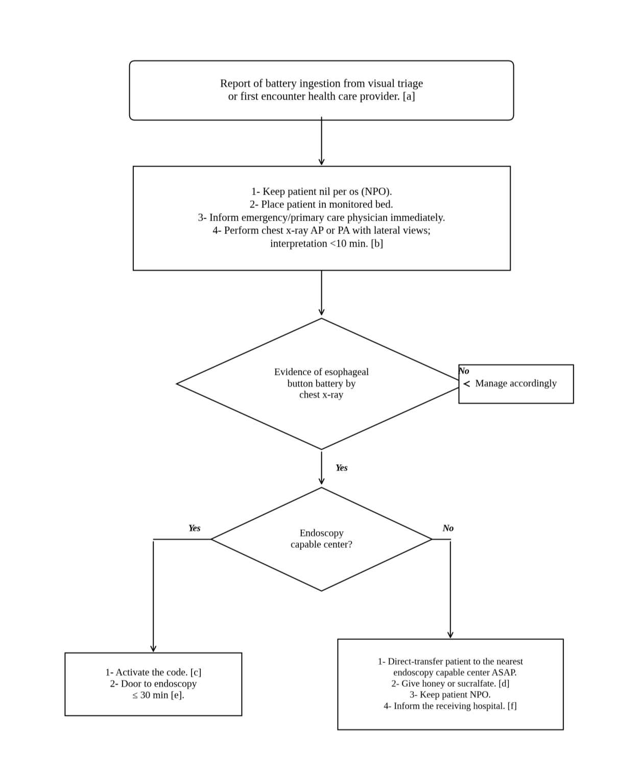
ACT scan is not typically required for every case of oesophageal button battery impaction. How- ever, it is essential when there is a significant risk of severe injury, particularly in cases of delayed presentation (more than 12 hours) or when there is clinical suspicion of complications [8,9]. Moreo- ver, the available medical literature supports prompt administration of honey or sucralfate as a temporizing intervention for oesophageal button battery ingestion. This approach is intended to mit- igate the severity of injury while awaiting defini- tive endoscopic removal. Optimal outcomes are observed when these treatments are rendered within 12 hours of ingestion and in the absence of perforation [10-14]. It is in light of such results that we propose the But- ton Battery Code (Appendix 1) to standardize management across institutions, in a similar man- ner to Trauma, ST-Elevation Myocardial Infarc- tion (STEMI), and Stroke codes. As demonstrated by this discussion, the duration between button battery ingestion and removal represents a critical, potentially modifiable determinant of injury sever- ity and adverse outcomes. Immediate identifica- tion and escalation are imperative, as tissue injury can begin within minutes and the risk for life-
threatening complications increases with each hour of continued exposure. The proposed <30- minute door-to-endoscopy target serves as an as- pirational benchmark to highlight current system delays and guide rapid activation, analogous in structure to other emergency response codes. The effects of individual adjunctive therapies (steroids, proton pump inhibitors, antibiotics) were not ana- lysed, as evaluating the relative contribution of these interventions was outside the scope of this study. While implementation of the Code may be chal- lenged by factors such as delayed recognition and limited endoscopic availability, the proposed pro- tocol is intended to standardize early recognition and care escalation to reduce management delays. Its effect on patient outcomes requires prospective evaluation, which should focus on measurable outcomes including time to endoscopy, success of initial removal, complication rates, and need for care escalation. In addition, standardized data col- lection would enable more precise assessment of the roles of adjunctive therapies and communica- tion strategies, as well as the identification of insti- tutional barriers to timely interventions. Such work is essential to translate recognition of de- layed management into measurable improve- ments in patient safety and outcomes.