Full text rendered from the published PDF. The PDF is the version of record; if the two differ, the PDF governs.
Gerota's fascia was exposed by dissection of the mobilised abdominal part of a 60-year-old formalin-fixed female cadaver during a routine anatomy dissection laboratory session, using Grant's Dissector Manual as reference, with digital images taken of the dissected area. The kidneys are paired retroperitoneal organs lying in the renal fossa on the posterior abdominal wall at the level of the T12 to L3 vertebrae, surrounded by three layers together with a partial fourth: the fibrous capsule attached directly to the kidney, the perinephric fat or adipose capsule, and the renal fascia, or Gerota's fascia, formed of collagenous connective tissue. Gerota's fascia fuses laterally and medially to the body of the kidney and superiorly to the adrenal glands, but is not fused inferiorly, so fluid may accumulate down the flank of the kidney without being present in the surrounding perinephric space. The fascia separates the kidney from the abdominal aorta, the inferior vena cava and the renal vessels. The article also addresses a persistent terminological confusion in which the terms "Gerota's fascia" and "Zuckerkandl's fascia" have each been applied to both the anterior and the posterior pararenal fascia. Reviewing the original sources, Chesbrough and colleagues found that Zuckerkandl characterised the posterior renal fascia in 1883 without reference to an anterior renal fascia, and that Gerota in 1895 assigned Zuckerkandl's name to the posterior renal fascia while confirming the existence of the anterior. The dissection images provide a gross anatomical reference for this structure.
Keywords: Gerota's Fascia, Zuckerkandl fascia, Renal fascia, Anatomy dissection, Clinical image
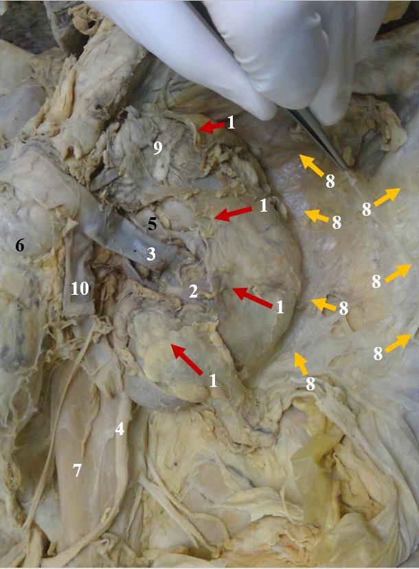
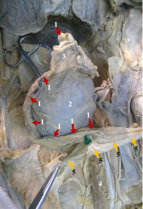
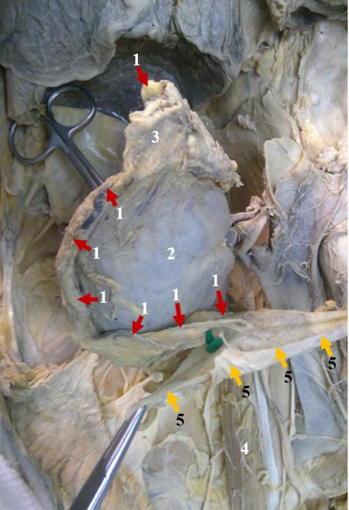
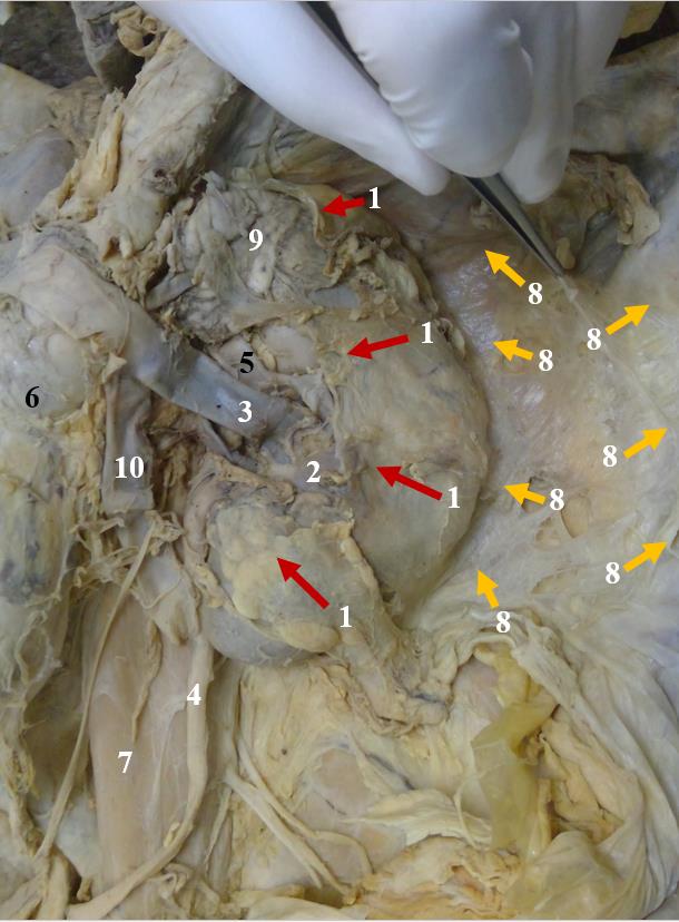
A mobilised abdominal part from a 60-year-old, formalin- fixed female cadaver was dissected during a routine anatomy dissection laboratory session. Grant’s Dissector Manual was used as a reference during the dissection process, and digital images were taken of the entire dissected area.
The kidneys are paired retroperitoneal organs situated in the renal fossa, on the posterior abdominal wall level with the T12 to L3 vertebrae. Each kidney is surrounded by three layers, as well as a partial fourth layer that furnishes a bed of fat posterior to each kidney [1].
The first layer, known as the fibrous capsule, attaches directly to the body of the kidney. The second layer is the perinephric fat capsule, or adipose capsule. The third layer, of most concern to us in this article, is the renal fascia or Gerota’s fascia, which is made up of collagenous connective tissue [2].
Gerota’s fascia surrounds the kidney, fusing laterally and medially to the body of the kidney, and superiorly to the adrenal glands. It is not fused inferiorly, however; this means that fluid may accumulate down the flank of the kidney whilst not being present in the perinephric space surrounding the kidney [3]. The significance of this fascia is that it separates the kidneys from other structures such as the abdominal aorta, inferior vena cava, and renal arteries and veins.
A common misunderstanding exists due to the frequent use, in medical literature, of the term “Gerota’s fascia” to describe both the anterior and posterior pararenal fascia. To further compound this misunderstanding, the term “Zuckerkandl’s fascia” has also been used to refer to either the anterior or posterior fascia. To clear up the confusion, Chesbrough RM et al. reviewed the original papers of both Gerota and Zuckerkandl.
They discovered that, while Zuckerkandl characterised the posterior renal fascia in 1883, his study made no mention of the anterior renal fascia. In 1895, Gerota firmly assigned Zuckerkandl’s name to the posterior renal fascia, while also confirming the existence of the anterior renal fascia [4]. Gerota’s research has led to the interchangeable use of the terms “Zuckerkandl’s fascia” and “posterior renal fascia” to describe the same anatomical structure. Likewise, the terms
Ahmad Dawalibi, Mohamad Bakir are with the College of Medicine, Alfaisal University, Riyadh, Saudi Arabia, e-mail: ahmad.dawalibi@hotmail.com, e- mail: mo7ammedbakir@gmail.com (Corresponding author). Ayman Behiery is with the Department of Anatomy, Alfaisal University, Riyadh, e-mail: abehiery@alfaisal.edu. DOI: 10.52609/jmlph.v3i3.81
“Gerota’s fascia” and “anterior renal fascia”, which refer to the same fascial layer, are also interchangeable [4].
Lastly, the outermost — what may be considered a fourth — layer, is known as paranephric fat and acts like a bed for the kidneys to lie on. It is present at the posterior and lateral sides of each kidney between the psoas major muscle and the quadratus lumborum muscle, isolating the kidneys from the muscles of the abdominal wall.
The integrity of the renal fascia is crucial for the detection of radiological abnormalities such as internal or subcapsular haematomas or abscesses. If the renal fascia is destroyed or the radiologist lacks the correct orientation, these anomalies may be ignored, potentially leading to missed diagnoses.
Our aim with these images is to illustrate Gerota’s fascia in a cadaveric kidney, as this is not well documented.
A genuine, gross image of Gerota’s fascia is extremely difficult to come by; instead, our main findings were sketchy photographs found on the internet. Thus, we firmly believe that a gross image obtained by cadaver dissection will represent a significant resource for medical students and physicians across various specialties. It will provide an accurate visual representation, allowing them to become familiar with the ap- pearance of the fascia from both a practical and an educational perspective.
Gerota’s Fascia, like any other fascia in the body, derives its significance from being responsible for, and related to, the spaces around it, which are clinically important with regard to the spread of fluids (blood in the case of trauma, exudate and pus in the case of infection, and metastasis in the case of tumours).
In the context of nephroblastomas and radical uretero- nephrectomy surgery, Gerota’s fascia and Zuckerkandl’s fascia are crucial. When treating nephroblastomas, the degree of perirenal infiltration is unknown; this necessitates the removal of a healthy tissue rim, including fibrous tissue and fat, along with the tumour, to achieve clear margins [5]. When performing a radical uretero-nephrectomy, the intact Gerota’s and Zuckerkandl’s fascia surrounding the kidney must also be removed. “Perifascial nephrectomy” is the medical term for this surgical technique [5]. To guarantee total tumour removal, achieve clean margins surrounding the kidney, lower the likelihood of tumour recurrence, and enhance patient outcomes generally, Gerota’s and Zuckerkandl’s fascia must be removed [5].
APPENDIX : Access the video and visually explore these structures by following the link provided: [https://youtu.be/EOR3c8mn5fQ]
The anatomy dissection laboratory session was held at Al- Faisal University’s College of Medicine in Riyadh, Saudi Arabia.
1. Soriano RM, Penfold D, Leslie SW. Anatomy, Abdomen and Pelvis: Kidneys. 2022 Jul 25. In: StatPearls [Internet]. Treasure Island (FL): StatPearls Publishing; 2023 Jan–. PMID: 29494007. 2. Marx WJ, Patel SK. Renal fascia: its radiographic im- portance. Urology. 1979 Jan;13(1):1-7. doi: 10.1016/0090- 4295(79)90002-5. PMID: 442312. 3. Okafor CN, Onyeaso EE. Perinephric Abscess. 2022 Aug 22. In: StatPearls [Internet]. Treasure Island (FL): StatPearls Publishing; 2023 Jan–. PMID: 30725621.
4. Chesbrough RM, Burkhard TK, Martinez AJ, Burks DD. Gerota versus Zuckerkandl: the renal fascia revis- ited. Radiology. 1989 Dec;173(3):845-6. doi: 10.1148/radiol- ogy.173.3.2682777. PMID: 2682777.
5. Tröbs RB. Anatomical basis for Wilms tumor surgery. Apr;14(2):50 2009 J Indian Assoc Pediatr Surg. - doi: 4. 10.4103/0971-9261.55151. 20671845; PMID: PMCID: PMC2905530.